| |
Research Tools |
|
| |
Sum Frequency Generation
Second Harmonic Generation
Molecular Biology
Tissue Culture
Surface Science
Spectroscopy & Chromatography
| |
|
Sum Frequency Generation |
|
| |
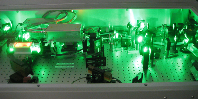
The vibrational sum frequency generation (VSFG) system consists of a Ti:sapphire oscillator as the seed laser and two Nd:YLF lasers as the pump for a regenerative amplifier. The regenerative amplifier gives a 6-W output at a repetition rate of 5 kHz. Half of the 6-W output is used to pump an optical parametric generator to generate an infrared beam.
VSFG spectroscopy provides molecule-level evidence concerning interfacial structures. We aim at exploring the potential of VSFG spectroscopy in the field of membrane biophysics. In particular, we focus on amyloid formation on membrane surfaces and cellular signaling through GPCR cross membranes.
Back to the top
|
|
|
Second Harmonic Generation |
|
| |
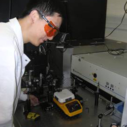
The Second Harmonic Generation (SHG) system utilizes either the 800-nm output from a Ti:sapphire oscillator or the UV-Visible output from an optical parametric generator pumped by the 6-W regenerative amplifier as the fundamental beam. The SHG system is set up to study both colloidal surfaces and planar surfaces.
We use the SHG system to investigate the surfaces of liposome bilayers and supported bilayers on solid substrates. We apply SHG to probe molecular interactions of biomolecules at membrane surfaces and kinetics of molecular transport across liposome bilayers.
Back to the top
|
|
| |
Molecular Biology |
|
| |
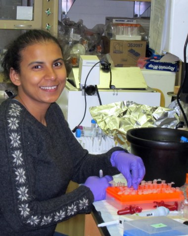
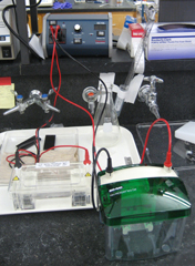
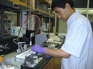
Our molecular biology lab allows us to prepare DNA constructs for protein expression in mammalian cells cultures. We perform DNA purification, PCR reactions, electrophoresis of DNA and proteins that enable us to clone and mutate genes of interest for biophysical studies.
We focus on making the DNA constructs for expressing membrane proteins and GPCR rhodopsin mutants. We also couple techniques of molecular biology to techniques of chemical biology to in vivo incorporate unnatural amino acids to proteins that can be further derivatized to spectroscopic tags to investigate conformational changes of proteins.
Back to the top
|
|
| |
Tissue Culture |
|
| |
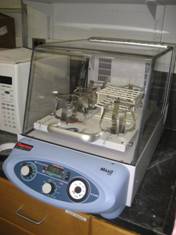
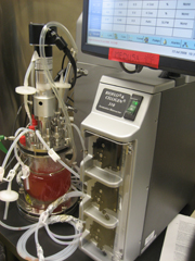
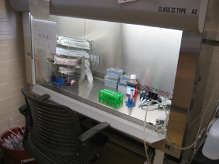
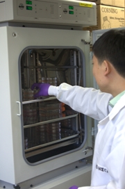
Our tissue culture lab is equipped to grow mammalian cells. These cells can be cultured on dishes in an incubator or in suspensions in a bioreactor to express recombinant proteins.
We use mammalian cells to express membrane proteins to ensure proper folding of transmembrane structures. The cells can be cultured in the bioreactor* that allows expression of proteins in large quantity for spectroscopic studies. The mammalian cells also serve as expression hosts to in vivo incorporate unnatural amino acids for tagging spectroscopic probe at specific sites of proteins.
*Medusa is the monstrous gorgon of Greek mythology who was cursed with snakes for hair. Our alumnus, Aditi Bhagat, named our bioreactor after Medusa because it has numerous plastic tubes protruding from the top and it is quite temperamental at times.
Back to the top
|
|
| |
Surface Science |
|
| |
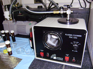
We set up the Langmuir-Blodgett trough to make and characterize model systems of cell membranes. Aside from liposome bilayers, we use supported lipid bilayers and lipid Langmuir monolayers as model systems of cell membranes. Using the Langmuir-Blodgett trough, we measure isotherms of lipid monolayers and study phase transition of lipid molecules at the air-water interface. We also use the trough to deposit lipid bilayers on solid substrates. The lipid monolayers and supported bilayers serve as platforms in the experiments of second-harmonic generation and sum frequency generation for probing electronic and vibrational structures of biomolecules at membrane surfaces.
Back to the top
|
|
| |
Spectroscopy & Chromatography |
|
| |
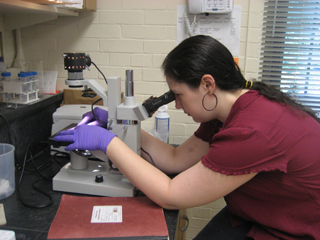
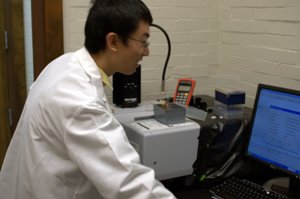
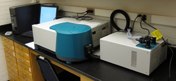
Our laboratory is equipped to carry out a number of chromatographic analyses of both protein and small-molecule samples. Our size-exclusion chromatography system allows purification and characterization of proteins and macromolecular assemblies such as nanodiscs. We use an HPLC chromatograph to measure the amount and conformation of small molecules such as retinal in biological samples.
We also use UV-visible and fluorescence spectroscopies to characterize our protein samples and perform functional assays. We employ UV-visible spectroscopy to study the activity of proteins such as the rod and cone visual pigments. We also perform both static and kinetic fluorescence measurements on multiple purified protein samples in order to track conformational changes in G-protein coupled receptors under physiological conditions.
Back to the top
|
|
| |
|
|
| |
Updated Mar. 2013 |
|